Radiology/Ultrasound
Radiology and ultrasound are valuable diagnostic tools used to evaluate a wide range of medical conditions in cats and dogs. These imaging services allow our veterinary team to look beyond what can be seen during a physical exam and gain important insight into your pet’s internal health.
Radiographs, commonly referred to as X-rays, are used to assess bones, joints, and internal organs. They can help identify fractures, foreign objects, arthritis, and changes within the chest or abdomen. X-rays are often quick to perform and can provide timely information to support diagnosis and treatment planning.
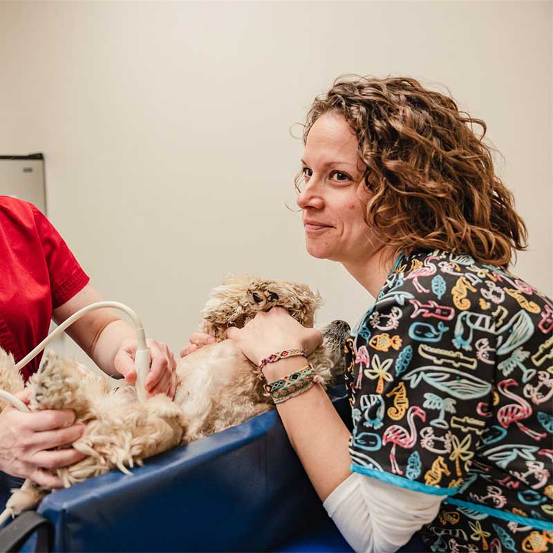
Ultrasound uses sound waves to create detailed images of soft tissues and organs such as the liver, kidneys, heart, bladder, and spleen. This imaging technique is especially helpful for evaluating internal organ disease, detecting masses or fluid buildup, and investigating conditions that may not be immediately apparent on examination. Ultrasound is noninvasive and generally well tolerated by pets.
Radiology and ultrasound are often used in combination with other diagnostic tools, including physical examinations and laboratory testing, to provide a more complete picture of your pet’s health. In addition to helping diagnose new conditions, these imaging services can also be used to monitor existing medical issues over time. Follow-up imaging may be recommended to track disease progression, evaluate response to treatment, or identify changes that require adjustments in care. This is particularly important in cases such as cancer, where monitoring the size or appearance of tumors can help guide ongoing treatment decisions.
By providing detailed information that cannot be obtained through a physical exam alone, radiology and ultrasound play a critical role in accurate diagnosis, treatment planning, and ongoing management of many medical conditions. These tools allow our veterinary team to deliver timely, informed care tailored to each pet’s needs.